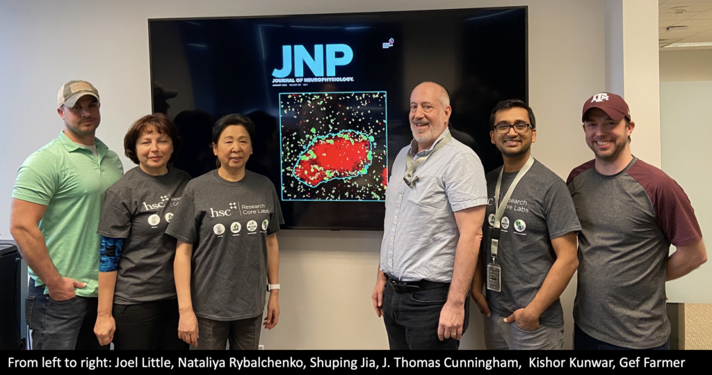
We are thrilled to find out confocal image taken in the Microscopy Core by Shuping Jia from the Cunningham lab and Kishor Kunwar from the Microscopy Core is being featured on the cover of the Journal of Neurophysiology. This is a remarkable achievement and a testament to these researchers' hard work and dedication.
Continue reading "Image taken in Microscopy Core featured on Journal of Neurophysiology cover page"Category Archives: Microscopy
Microscopy Core adds Huygens-Image processing and analysis software to its toolkit
Huygens Essential is a powerful imaging software that allows users to process and analyze large sets of microscopy data. Developed by Scientific Volume Imaging, it is compatible with a wide range of microscope platforms, including widefield, confocal, and super-resolution systems. Huygens Essential offers a wide range of advanced image processing tools, including deconvolution, registration, and segmentation. It also provides support for a variety of file formats, including image stacks, multi-dimensional datasets, and time-lapse sequences.
Continue reading "Microscopy Core adds Huygens-Image processing and analysis software to its toolkit"Zeiss Axioscan 7 coming to Histology Core
Histology Core will be equipped with Zeiss Axioscan 7 automated slide scanner. This powerful imaging equipment allows for high-resolution digital scanning of tissue samples at an unparalleled speed.
Continue reading "Zeiss Axioscan 7 coming to Histology Core"